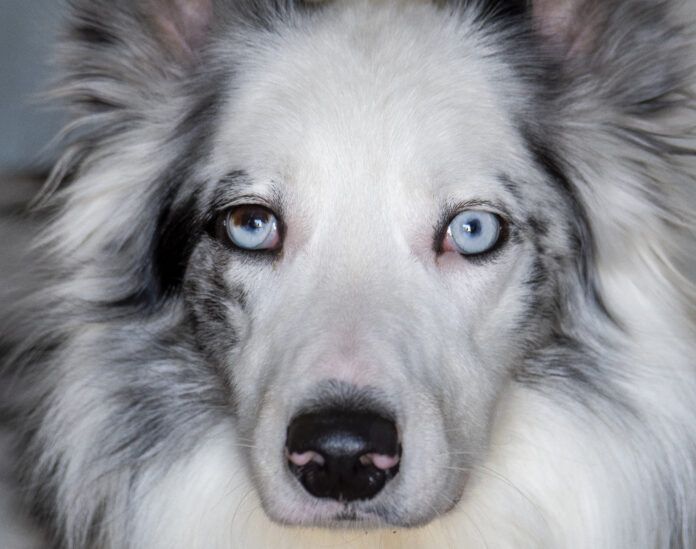
Bumping into furniture, reluctance to go outside at night—these are signs of visual impairment. There are several causes of visual impairment and blindness in dogs—one of those causes is progressive retinal atrophy (PRA).
PRA is the gradual destruction of photoreceptors in the retina. The retina is located at the back of the eye. Photoreceptors in the retina capture light that enters the eye and convert the light into electrical signals. These signals are sent to the brain through the optic nerve. The brain converts these electrical signals into an image of what your dog sees.
There are two types of photoreceptors in the retina—rods and cones. Rods are responsible for detecting the level of light (or brightness) and motion. Cones are responsible for detecting color. Unlike humans, dogs only have two types of cones and can perceive the colors blue and yellow but not red and green.
PRA begins with the loss of rods in the retina. This causes dogs to develop night blindness, also known as nyctalopia. Dogs with nyctalopia may initially exhibit reluctance to go outside after dark. As more rods are destroyed, these dogs will develop difficulty tracking moving objects in dim light and then in brighter light.
PRA will progress from destruction of rods to loss of cones in the retina. As cones are destroyed, dogs with PRA will gradually lose visual acuity and eventually become blind. Since their vision loss is gradual, these dogs will have created a mental map of their environment. They may not show symptoms of visual impairment until they are introduced to a new environment (like a new home) or if their current home is remodeled or the furniture rearranged.
Causes of Progressive Retinal Atrophy in Dogs
PRA is an inherited condition in dogs. Certain breeds of dogs are more likely to inherit the gene mutations necessary to cause PRA. These include (but are not limited to) Poodles, Labrador Retrievers, Golden Retrievers, Irish Setters, and Shetland Sheepdogs.
You can have your dog tested for the gene mutations that are known to be associated with the development of PRA. The American Kennel Club (AKC) offers DNA testing for PRA and other inherited conditions through Neogen. The University of California at Davis (UC Davis) offers testing for the PRA gene mutations of specific breeds. Both laboratories complete testing on a cheek swab sample that you can collect at home and submit by mail to the laboratory.
Testing your dog for the gene mutations that are associated with development of PRA will give you more information about whether your dog is likely to develop PRA. Dogs that possess the necessary gene mutations should undergo an annual examination with a veterinary ophthalmologist to screen for development of PRA.
Not all of the gene mutations associated with developing PRA have been identified. Research into the genetics of PRA is ongoing and our knowledge of the gene mutations behind this condition continues to grow.
Diagnosing PRA in Dogs
Proper diagnosis of PRA in your dog will require a visit to a veterinary ophthalmologist. Your dog will receive a complete ophthalmologic exam, including an electroretinogram (ERG). An ERG records the electrical activity of the retina when exposed to light. Dogs with PRA will have characteristic changes to their retinal electrical activity due to the destruction of photoreceptors in the retina.
The ERG takes about 20 to 30 minutes to complete. Your dog is first placed in a completely dark room for about 15 minutes. This immersion into darkness makes the retina particularly sensitive to light.
The pupils will need to be dilated for this test. If your dog’s pupils are not already dilated, a drop of a pupil dilation solution (such as tropicamide ophthalmic solution) will be instilled in each eye. Then a drop of a cornea numbing solution (such as proparacaine or tetracaine) is applied to the surface of each eye. This numbs the surface of the cornea in preparation for the ERG.
A contact lens that contains an electrode is applied to the surface of your dog’s cornea. Two electrodes are applied to your dog’s skin. One electrode is placed at the top of his skull and the other is placed next to the eye being tested.
Pulses of light are shone into the eye being tested. Electrical activity of the retina is recorded by the electrodes and the output is displayed on a computer. Characteristic changes in the retinal electrical activity confirm the diagnosis of PRA.
Your dog needs to sit still without moving his head during the ERG. Most dogs will require a sedative for the procedure. Some dogs may need to be briefly anesthetized to facilitate completion of the ERG.
Treatment and Prognosis for Dogs with PRA
There is no way to prevent the development of PRA in dogs. All dogs that have been diagnosed with PRA will lose their sight. PRA is not a painful condition. Most dogs will adjust to being visually impaired and lead a full and happy life.
A supplement called Ocu-GLO may have a protective effect on the retina. Anecdotal evidence appears to indicate that Ocu-GLO may slow the progression of PRA, although more research is necessary to prove this effect. Ocu-GLO and other eye supplements will not stop dogs from going blind due to PRA.
Dogs with PRA are at increased risk for developing cataracts. Cataracts are a clouding of the lens in the middle of the eye. Molecules released by the photoreceptors as they degenerate can be toxic to the lens and cause a cataract to develop.
Cataracts can cause proteins to leak from the damaged lens. These proteins can cause inflammation of the inner lining of the eye—this is called uveitis. Uveitis can cause an increase in the intraocular pressure of the eye—this is called glaucoma. Glaucoma is a painful condition that can be treated if it is detected early.
Some breeds that are at increased risk of developing PRA are also at risk for developing hereditary cataracts. Like PRA, hereditary cataracts occur in dogs that have inherited the gene mutations that cause this condition. Breeds that are genetically predisposed to developing PRA and hereditary cataracts include (but are not limited to) the American Cocker Spaniel, Labrador Retriever, and Australian Shepherd.
Dogs that have been diagnosed with PRA should undergo regular ophthalmologic exams with their primary care veterinarian to screen for the development of cataracts and glaucoma. This exam should include a test of the intraocular pressure in each eye. Intraocular pressures are measured with a device called a tonometer. If your dog’s intraocular pressures are high, then your veterinarian will prescribe one or more eye medications to treat your dog’s glaucoma.
Most dogs with PRA will gradually lose their vision and become blind over a period of one to two years. The speed at which your dog will become blind from PRA will depend on his breed and the underlying genetic cause of his condition.


