Progressive Retinal Atrophy (PRA) in Dogs
There are several causes of visual impairment and blindness in dogs—one of those causes is progressive retinal atrophy (PRA). PRA is the gradual destruction of photoreceptors in the retina.
Pannus in Dogs
Pannus is a growth of vascular tissue over the cornea. It usually starts on the edges of the cornea and spreads into the center of the eye. Untreated, it can coat the entire cornea, which is how it causes blindness.
Dog Eye Allergies vs. Infections
Deciding if your dog has an allergic eye reaction or if he is starting with an infection is difficult. Unfortunately, eye problems can go from minor to very serious very quickly.
Tricks to Giving Your Dog Eye Medication
Eye drops and ointments are prescribed for a variety of ocular conditions in dogs, including glaucoma, corneal ulcers, and recovery from cataract surgery. Even a minor eye infection like conjunctivitis what the kids in your grade-school days called pink eye" can require regular administration of drops."
Lenticular Sclerosis in Dogs
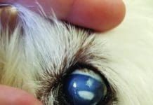
Lenticular sclerosis in dogs is a normal aging change in the dog’s lenses that results in that bluish haze. It is not painful, and it will never make your dog blind.
Are Dogs Colorblind?
A dog’s world isn’t all black, gray, and white. Much like a colorblind person, dogs can see color, but some colors like red and green are not distinct to a dog.
Dog Tear Stain Remover: When and What to Use
Dog tear stain removers can remove tear stains. However, tear stains are harmless when they aren't caused by another issue and changes in diet and grooming can also resolve dog tear stains.
Tips on Living With and Training a Blind Dog
When Orbit came into one of the classes I offer for puppies and their owners, in Santa Cruz, California, he was in most ways...
Entropion in Dogs: How to Treat This Common Eye Problem
This Labrador has acquired entropion, secondary to dehydration and renal disease. It should resolve somewhat once he is rehydrated.üThis dog has severe, 360-degree entropion (meaning the eye is being touched by the upper and lower eyelids, and is irritated from all sides). Notice the tearing and matting around the eye from irritation, as well as the prominence of the third eyelid, which has elevated in order to protect the cornea.üHere is the same dog is after surgical repair for his severe entropion. The eye is clear and bright, without evidence of matting or squinting. (The greenish-yellow color is fluorescein eye stain. This is a test that uses a dye and a blue light to detect foreign bodies or damage to the cornea. Happily, this dog had neither!)
Cloudy Eyes in Dogs
There are many possible ways in a which a dog's eyes can look clouded. Often, you are seeing the cloudiness in the lens of the eye an elastic, transparent structure that lies behind the iris (the pigmented part of the eye) and the pupil (the opening in the center of the eye). Tiny muscle fibers inside the eye contract and relax to makes the lens change thickness and shape; these movements help the dog change focus. As dogs age, certain changes cause the lens to turn white and become visible. When this ordinarily transparent structure develops a cloudy spot or section, the dog's vision is compromised.
Structure of the Canine Eye
The dog's eye is pretty much a garden-variety mammalian eye, with some notable adaptations that have evolved over the millennia. It is a globe with two fluid-filled chambers (anterior and posterior). The chambers are separated by the lens, the structure that helps focus light beams onto the rear part of the eye, the retina. The eye's outer, clear surface, the cornea, offers protection to the inner eye and helps the lens focus light onto the rear of the eyeball, the retina.
Older Dogs and the Onset of Cataracts
Cataracts make the lens of the eye opaque or cloudy, which gradually reduces vision to the point of blindness. In their early stages, cataracts cause blurring and distortion of vision, but they are invisible to the naked eye. By the time most owners notice them, cataracts involve more than 60 percent of the dog's eye. Cataracts often accompany other illnesses, such as diabetes and hypothyroidism (low thyroid function). Surgery performed by a veterinary ophthalmologist is the only treatment considered effective in conventional veterinary medicine and is indicated only in cases where the cataracts are not a result of a secondary disease such as diabetes.


